Microscopy can reveal a Titanium electrode coating's visible structure, local damage and coverage at sampled locations, but cannot establish operating life by itself. You receive two dramatic close-ups from competing suppliers and neither identifies where those images came from. Choosing on appearance alone leaves your team comparing photographs instead of equivalent evidence. A defined sampling plan, matched imaging conditions and separate functional tests make that comparison usable.
1. What can Titanium electrode microscopy establish?
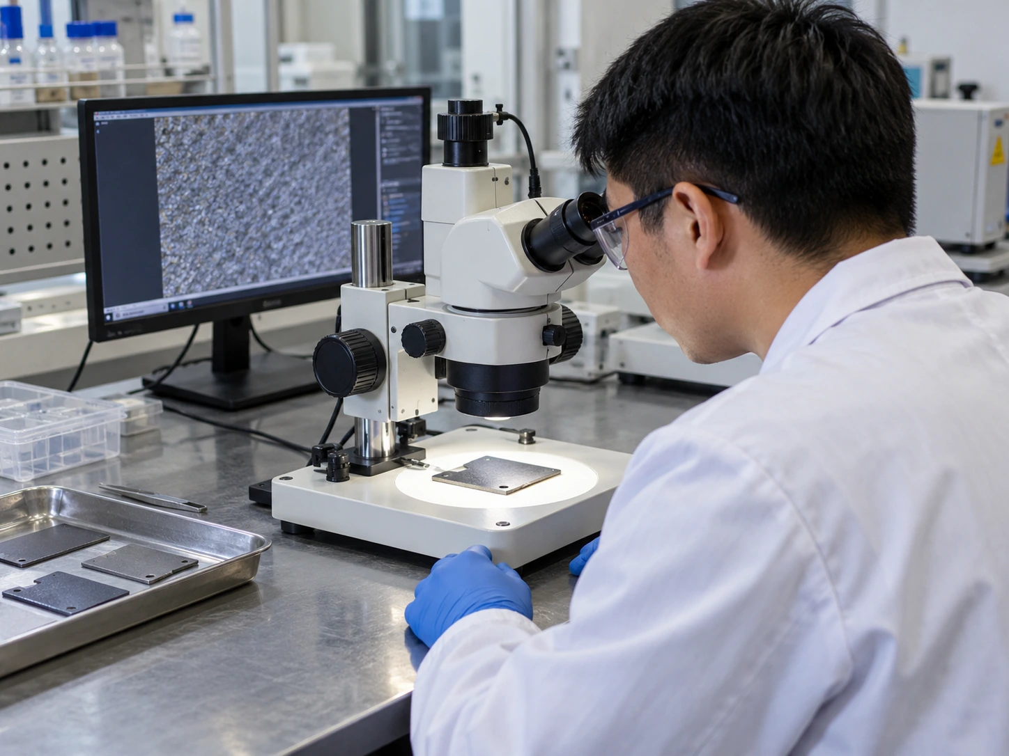
Microscopy establishes visible features within sampled areas of a Titanium electrode, including texture, deposits and exposed edges. Its conclusions stop at the resolution and locations examined.
Which decision should the image support?
Request an image to answer a specific question: surface damage, an unusual deposit or a change between lots. Start with that inspection question. A supplier can then propose the required view instead of sending an attractive but unrelated close-up.
- Identify the electrode face, position and condition before imaging.
- Describe the concern without assuming that an observed mark caused a failure.
| Check | Decision |
|---|---|
| Local surface concern | Map the location and compare adjacent areas |
| Whole-lot acceptance | Define representative sampling beyond one image |
A sharply focused field still covers only part of your component. Key Takeaway: Use microscopy to narrow a question before choosing a separate acceptance test.
2. Which Titanium electrode images answer your question?
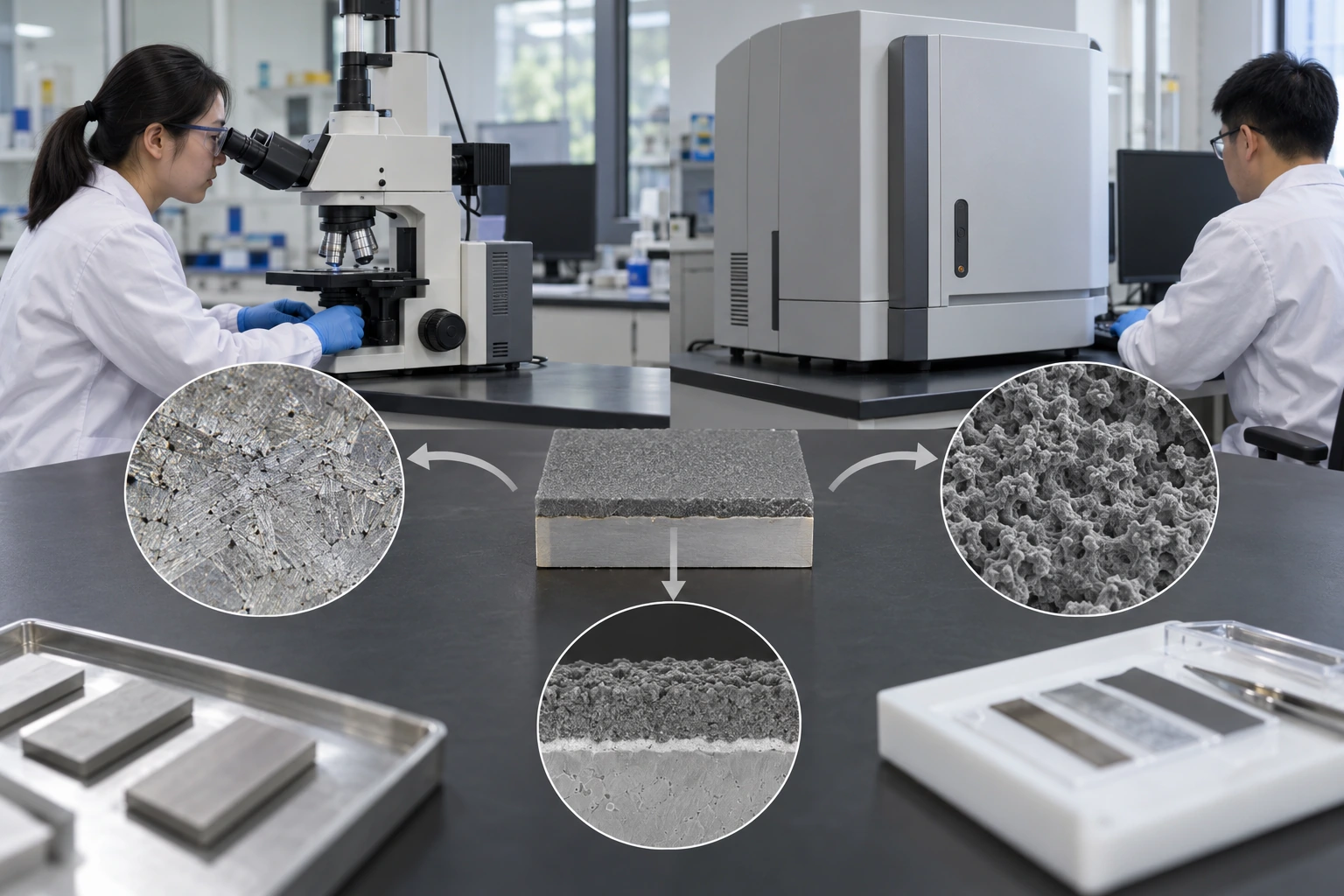
Choose optical or electron microscopy for a Titanium electrode according to feature size and the question being investigated. Neither technique becomes a chemical assay simply because its image looks detailed.
Do you need context or finer detail?
A low-magnification overview keeps a mark connected with its surroundings; finer views can then resolve smaller features. Keep those views together. An isolated close-up can hide whether an observation concerns a cut edge, a weld boundary or a coated working face.
- Retain a whole-part photograph before selecting magnified regions.
- Ask the laboratory to justify the instrument and detector for the required observation.
| Check | Decision |
|---|---|
| Overview | Shows position and extent across a larger area |
| Detailed view | Resolves local features within a smaller field |
Changing magnification changes the question an image can answer. Key Takeaway: Specify the needed feature scale instead of buying the largest magnification number.
3. How should Titanium electrode sampling be planned?
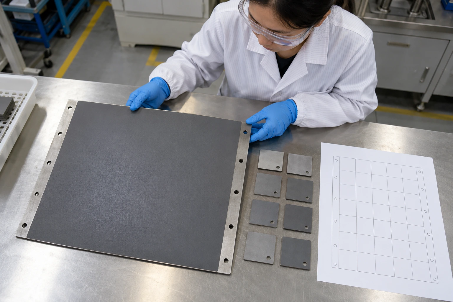
A Titanium electrode sampling plan should cover agreed locations and distinguish routine specimens from suspect areas. A deliberately selected defect image cannot describe the frequency of that defect across a batch.
How will you avoid cherry-picking?
Divide the component into functional regions before choosing locations, then retain that selection record. Include the ordinary areas too. If investigation requires extra images of a damaged spot, identify them as targeted evidence beside the planned sample set.
- Separate routine sampling from additional investigation of a suspect region.
- Record whether specimens came from finished parts, witness coupons or returned equipment.
| Check | Decision |
|---|---|
| Planned location | Supports a defined comparison across samples |
| Targeted anomaly | Describes that concern without establishing lot prevalence |
Sample selection determines what you can reasonably generalize. Key Takeaway: Agree on locations before either party sees the most persuasive photograph.
4. Why must Titanium electrode images retain their scale?
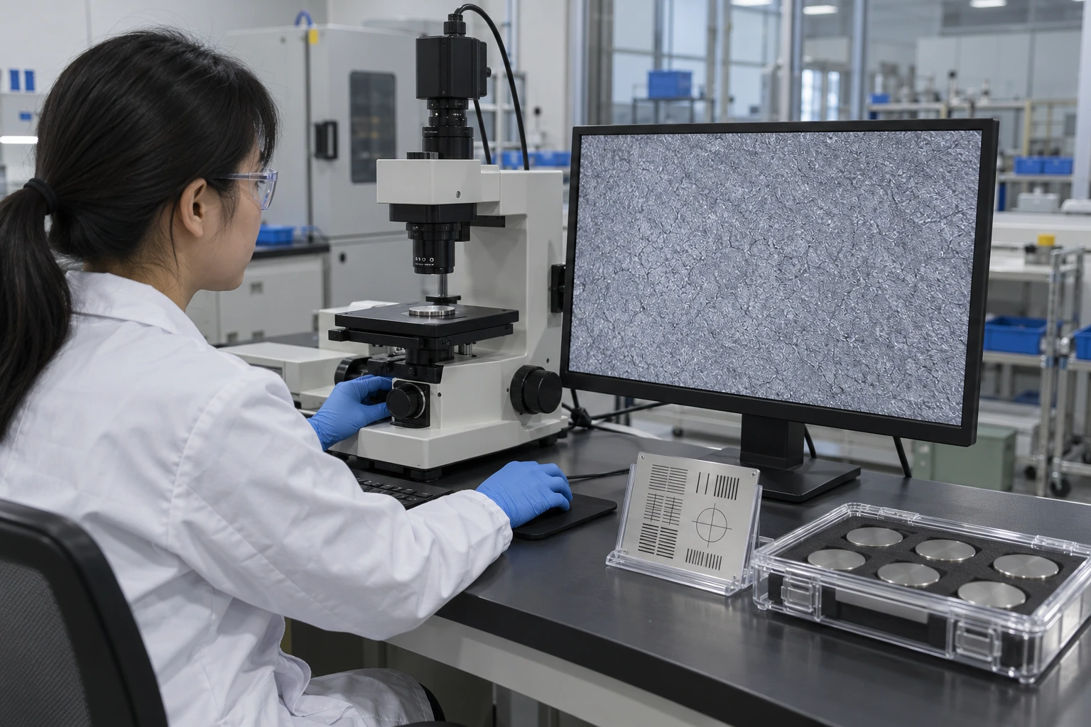
A Titanium electrode image needs a valid scale and acquisition record so you can compare features across reports. Resizing an image can make a printed magnification label misleading.
What belongs beside the original image?
Retain the original file, scale calibration, detector information and specimen orientation with each image. Preserve the measurement context. Include these requirements in a custom engineering review so drawing locations and microscopy locations use the same reference system.
- Keep original image files alongside presentation copies.
- Ask how scale was calibrated and whether any resizing or processing occurred.
| Check | Decision |
|---|---|
| Calibrated scale | Supports dimensional interpretation within stated limits |
| Untracked screenshot | Can support discussion but weakens measurement claims |
A visual comparison is easier to defend when its scale survives copying. Key Takeaway: Reject dimension claims based only on an enlarged screenshot without a calibration basis.
5. Do Titanium electrode surface cracks prove failure?
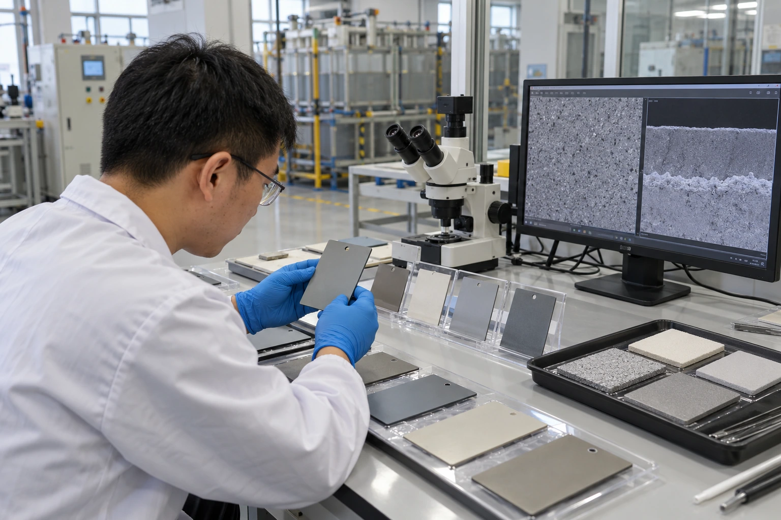
Surface cracks in a Titanium electrode oxide coating do not automatically prove failure, because some thermally prepared coatings have cracked surface morphologies. New damage, detachment and changes during service still warrant investigation.
What changed from the accepted condition?
Compare like-for-like images with an approved sample or a retained specimen from the same process. Look for a documented change. Crack appearance alone cannot tell you whether the coating remains attached, retains catalytic activity or meets an agreed service requirement.
- Distinguish fine surface morphology from visible flaking or newly exposed substrate.
- Compare equivalent locations and imaging conditions before attributing a difference to manufacturing.
| Check | Decision |
|---|---|
| Accepted baseline texture | Interpret within the qualified coating process |
| New localized damage | Investigate with handling and operating records |
A baseline reduces the chance of mistaking normal texture for a new defect. Key Takeaway: Escalate observed changes with evidence instead of judging every crack by appearance.
6. Can Titanium electrode microscopy confirm composition?
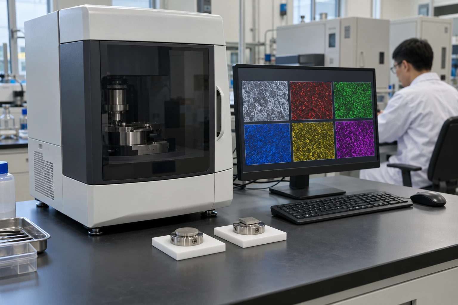
Microscopy alone cannot confirm a Titanium electrode coating's chemical composition; attached analytical methods provide different evidence. Elemental analysis also has sampling and preparation limits.
Are you comparing pictures or analytical results?
An elemental map can show where a detected element occurs within the examined region. Ask for the underlying measurement. Surface topography, coating thickness and the interaction volume can affect interpretation, so a colored image should not replace a documented analytical result.
- Identify which method generated a composition claim and which region it examined.
- Keep elemental identification separate from claims about chemical state or coating loading.
| Check | Decision |
|---|---|
| Surface image | Documents morphology under specified imaging conditions |
| Elemental analysis | Requires its own method, calibration and interpretation |
Different evidence types answer different material questions. Key Takeaway: Request chemistry data only when chemistry is part of your acceptance decision.
7. Can Titanium electrode cross-sections prove coverage?
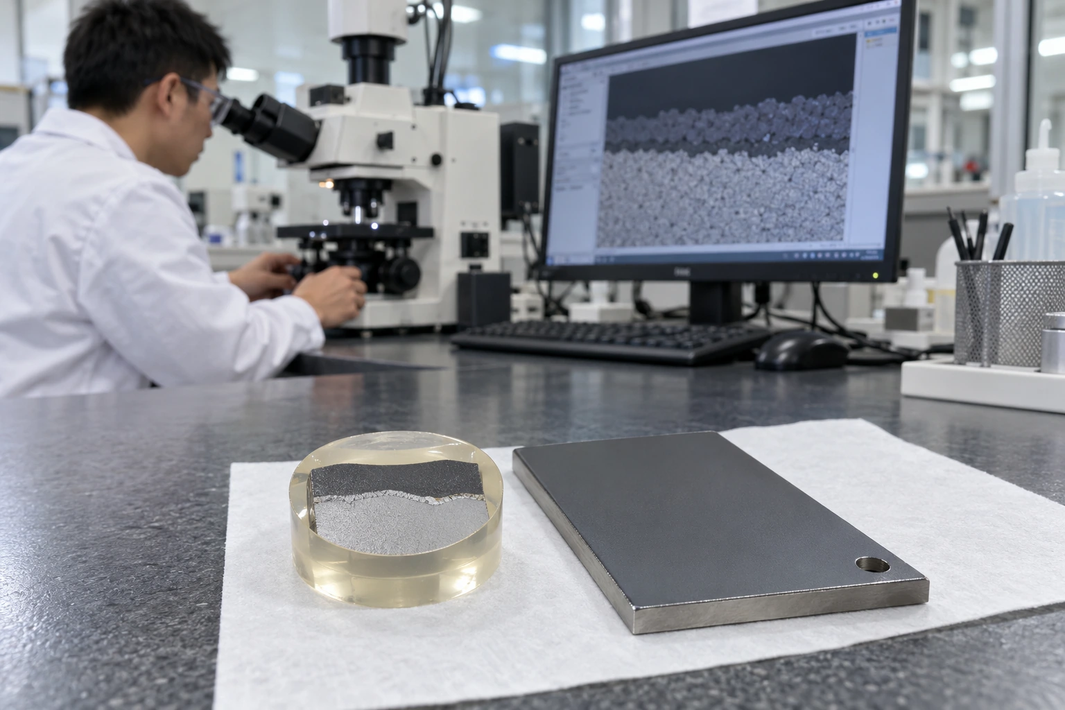
A Titanium electrode cross-section can reveal a local coating-substrate interface, but one cut cannot establish coverage across an entire electrode. Section preparation can also introduce marks that resemble damage.
Where should the section be taken?
Choose the cut location before destroying the specimen and retain an image of the intact surface. Connect the section with its origin. Ask the laboratory to distinguish preparation artifacts from features present before cutting, mounting or polishing.
- Approve destructive sampling locations and retain an untouched comparison specimen.
- Document section orientation and whether an edge or working face was selected.
| Check | Decision |
|---|---|
| Mapped cross-section | Provides localized interface evidence |
| Unlocated section | Cannot reliably represent a drawing region or whole part |
A section is strongest when you can trace it back to the original surface. Key Takeaway: Use cross-sections selectively where interface evidence changes your decision.
8. How can Titanium electrode preparation distort evidence?
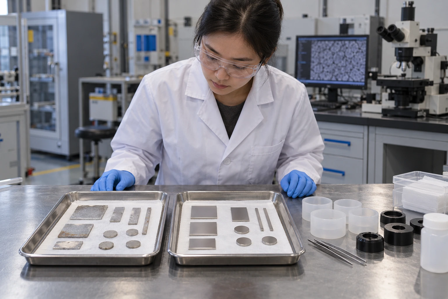
Preparation can alter a Titanium electrode specimen through cleaning, cutting, mounting or an added conductive coating. Record those operations before interpreting residue or surface chemistry.
Could preparation erase the evidence?
For a returned component, preserve an as-received record before cleaning or removing deposits. Keep the untreated reference available. This is especially useful when investigating water-treatment applications, where deposits and operating residues may help distinguish service exposure from a manufacturing concern.
- Separate as-received photographs from images taken after preparation.
- Record added mounting or conductive materials that could affect analysis.
| Check | Decision |
|---|---|
| As-received record | Preserves the initial condition and visible residues |
| Prepared specimen | Supports the chosen method with declared alterations |
Preparation improves access to some features while changing others. Key Takeaway: Agree on preservation and preparation before shipping an investigation sample.
9. Can Titanium electrode images predict working life?
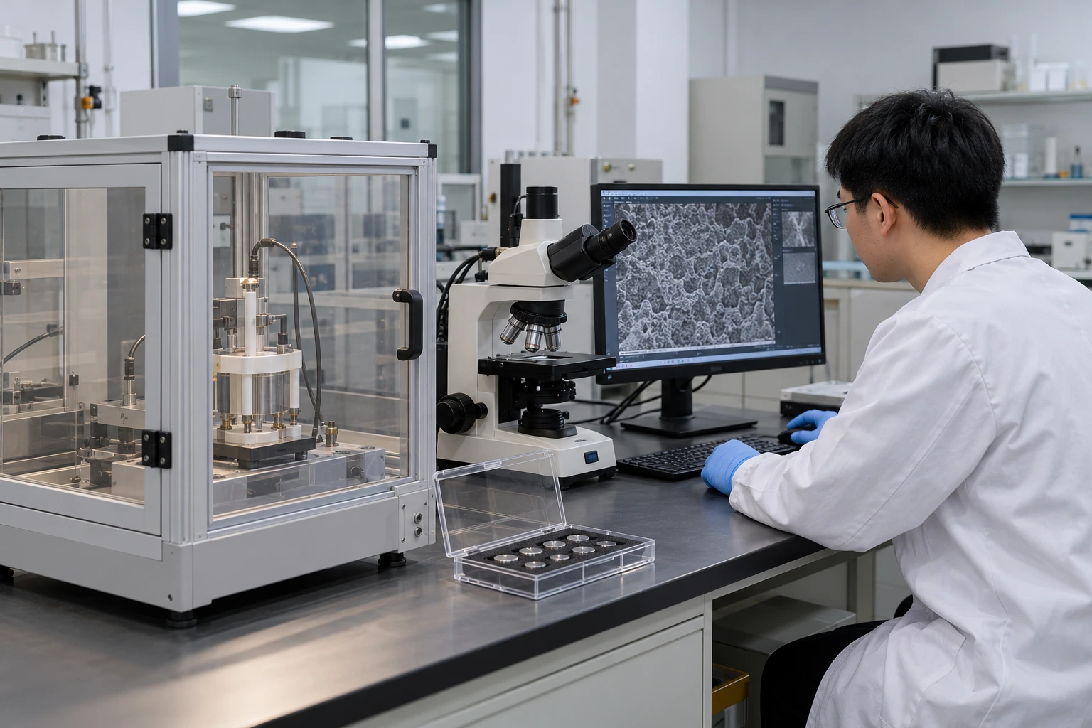
Images cannot predict a Titanium electrode working life without evidence linking the observed features to a defined operating duty. Service testing answers a different question from surface observation.
Which evidence belongs alongside the images?
Compare imaging results with relevant operating records, electrical behavior and any agreed functional test. Keep performance evidence separate. A coating can look similar while behaving differently, and a visible difference may not change performance within the tested duty.
- Record electrolyte, temperature and operating history for used samples.
- Avoid converting a surface feature into a lifespan claim without validated correlation.
| Check | Decision |
|---|---|
| Morphology record | Describes the examined structure |
| Duty-specific test | Evaluates behavior under declared operating conditions |
Combining evidence is useful only when the samples and test boundaries match. Key Takeaway: Approve an electrode using the agreed evidence package, not a photograph alone.
10. What Titanium electrode image evidence should you request?
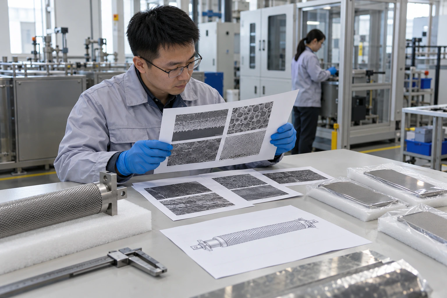
Request a Titanium electrode image package that identifies samples, locations, preparation, scale and the decision each image supports. This resolves ambiguous comparisons before they become purchasing disputes.
What should your next inquiry contain?
For drawing-based electrode selection, contact us today with your application, component drawing and the observation you need clarified. Define the inspection scope before quoting. Our position is that a useful electrode specification connects evidence with operating duty; any specialist laboratory work must be confirmed separately.
- Share the specific concern and any accepted reference condition.
- Confirm which inspection records are included and which specialist tests need a separate agreement.
| Check | Decision |
|---|---|
| Defined question | Allows a relevant inspection proposal |
| Defined acceptance basis | Makes supplier comparisons more consistent |
The review should end with a clear decision or a named unresolved question. Key Takeaway: Ask for evidence that changes your purchasing decision and retain the original records.
Frequently Asked Questions
Can I approve a coating from one SEM photograph? No. One field shows one sampled region; acceptance also needs an agreed sampling plan and relevant functional evidence.
What's the best magnification for electrode inspection? It depends on the feature. Keep an overview and let the required resolution determine the detailed imaging method.
How do I know if a dark patch is missing coating? Appearance alone cannot settle it. Compare imaging conditions and request a suitable analytical or cross-sectional check where needed.
Can I clean a returned electrode before sending it for analysis? Only after agreeing on the investigation plan. Cleaning can remove deposits or residues that help explain the observed condition.
What's the best way to compare two suppliers' images? Use a common sampling and imaging brief. Match locations, specimen preparation and scale before comparing surface features.